Let us know
Enter your information for the latest news
[wpforms id="13471"]
I consent to the processing of personal data and agree with the user agreement and privacy policy


Cancer is a complex and diverse group of diseases, with each type exhibiting unique molecular characteristics. Accurate diagnosis is crucial for selecting the most effective treatment options and improving patient outcomes. Among the diagnostic tools used in modern pathology, Immunohistochemistry (IHC) staining stands out as a vital technique for identifying specific cancer markers and differentiating between various types of cancer. IHC Stainer plays a pivotal role in cancer diagnosis, guiding therapeutic decisions and advancing research.
In this article, we will delve into the significance of IHC staining in cancer diagnosis, its underlying principles, advantages, and how it contributes to better treatment outcomes.
Immunohistochemistry (IHC) staining is a laboratory technique used to detect the presence of specific antigens or proteins in tissue samples. By utilizing antibodies that specifically bind to target proteins, IHC Stainer helps pathologists visualize the expression and localization of these proteins under a microscope. The process involves labeling the antibodies with a color-producing enzyme or fluorescent dye, allowing the targeted antigens to be easily identified within the tissue.
IHC staining is widely used in pathology for diagnosing various diseases, including cancer. The technique is particularly valuable for detecting protein markers associated with tumor cells, determining the cancer type, and assessing the tumor’s characteristics. Its applications extend to identifying hormone receptors, growth factors, and other biomarkers that are crucial in cancer treatment.
Cancer diagnosis often requires a detailed analysis of tissue samples to identify specific molecular features of the tumor. IHC staining is an essential tool in this process, offering insights that go beyond traditional histological examination. Here are some key roles of IHC staining in cancer diagnosis:
Differentiating Cancer Types
Not all cancers look the same under a microscope, making it challenging for pathologists to differentiate between similar-looking tumors. IHC Stainer helps distinguish between different cancer types by detecting specific protein markers that are uniquely expressed in certain cancers. For example, distinguishing between primary lung cancer and metastatic cancer originating from another organ can be challenging using only routine histology. IHC markers such as thyroid transcription factor-1 (TTF-1) can help confirm the diagnosis of lung cancer, while other markers may indicate metastasis from a different site.
Determining Tumor Origin
When a metastatic tumor is found, it is crucial to identify its tissue of origin. IHC staining can provide valuable information about the primary site by detecting organ-specific markers. For example, the presence of prostate-specific antigen (PSA) in a metastatic lesion strongly suggests that the primary cancer originated in the prostate. Similarly, markers such as cytokeratin 7 (CK7) and cytokeratin 20 (CK20) are used to help determine the site of origin for carcinomas, guiding further investigations and treatment.
Identifying Prognostic and Predictive Markers
IHC staining is also important for detecting biomarkers that have prognostic or predictive significance. Prognostic markers provide information about the likely course of the disease, while predictive markers indicate how likely a tumor is to respond to a specific treatment. For example, HER2/neu overexpression in breast cancer is associated with a more aggressive disease but also predicts a favorable response to targeted therapies such as trastuzumab. Similarly, detecting the expression of estrogen and progesterone receptors helps determine the suitability of hormone therapy for breast cancer patients.
Assessing Tumor Grade and Aggressiveness
Some IHC markers can indicate the aggressiveness of a tumor, providing valuable insights into the expected behavior of the cancer. Ki-67, a marker of cell proliferation, is commonly used to assess tumor grade in cancers such as breast, prostate, and neuroendocrine tumors. A higher Ki-67 index suggests rapid cell growth and a potentially more aggressive tumor, helping oncologists tailor the treatment plan accordingly.
Personalizing Cancer Treatment
Precision medicine aims to customize cancer treatment based on the individual characteristics of each patient’s tumor. IHC staining enables the identification of specific molecular targets that can be exploited for personalized therapy. For instance, immunotherapies targeting the programmed death-ligand 1 (PD-L1) pathway require testing for PD-L1 expression in the tumor to predict response to treatment. The ability to detect these markers through IHC facilitates the use of targeted therapies, immunotherapies, and other personalized approaches in cancer management.
IHC Stainer offers several advantages over traditional diagnostic techniques, making it an invaluable tool in cancer diagnosis. Here are some of the key benefits:
High Sensitivity and Specificity
IHC staining provides a highly sensitive and specific method for detecting proteins within tissues. The use of monoclonal antibodies, which are designed to bind to a single epitope on the target antigen, allows for precise identification of cancer markers. This accuracy is essential for reliable diagnosis and treatment planning.
Visualization of Protein Localization
Unlike other molecular techniques that detect proteins in solution, IHC Stainer allows for the visualization of protein expression within the context of the tissue architecture. This ability to see the exact location and distribution of proteins helps pathologists understand the interactions between cancer cells and their microenvironment, providing insights that are crucial for diagnosis and prognosis.
Cost-Effective and Accessible
IHC Stainer is relatively cost-effective compared to more advanced molecular diagnostic techniques, such as next-generation sequencing. The technique is widely available in pathology laboratories, making it an accessible option for most healthcare facilities.
Compatibility with Routine Pathology Workflows
IHC staining integrates seamlessly with standard histological procedures, allowing for simultaneous evaluation of tissue morphology and protein expression. This compatibility makes it a practical and efficient choice for routine cancer diagnosis.
Despite its many benefits, IHC staining does have some limitations that need to be considered:
Antibody Quality and Specificity
The quality and specificity of the antibodies used in IHC Stainer are critical for obtaining accurate results. Poor-quality antibodies or those with low specificity may result in false-positive or false-negative results, potentially leading to misdiagnosis.
Interpretation Variability
IHC staining results can be subjective, with variability in interpretation between pathologists. Standardization of staining procedures and scoring systems can help minimize discrepancies, but interpretation remains a challenge in some cases.
Limited Detection of Genetic Alterations
While IHC Stainer is effective for detecting protein expression, it cannot identify genetic mutations or alterations in the DNA sequence. Complementary molecular techniques, such as fluorescence in situ hybridization (FISH) or polymerase chain reaction (PCR), may be needed for a comprehensive cancer diagnosis.
Beyond its diagnostic applications, IHC staining is a valuable tool in cancer research. It allows researchers to study protein expression patterns in different types of tumors and understand the molecular mechanisms underlying cancer progression. IHC staining can also be used to evaluate the effectiveness of new therapeutic agents by examining their impact on specific protein markers in preclinical and clinical studies.
Furthermore, advances in IHC Stainer technology, such as multiplex IHC, enable the simultaneous detection of multiple protein markers within a single tissue section. This approach provides a more comprehensive view of the tumor microenvironment and helps identify complex interactions between cancer cells, immune cells, and other components.
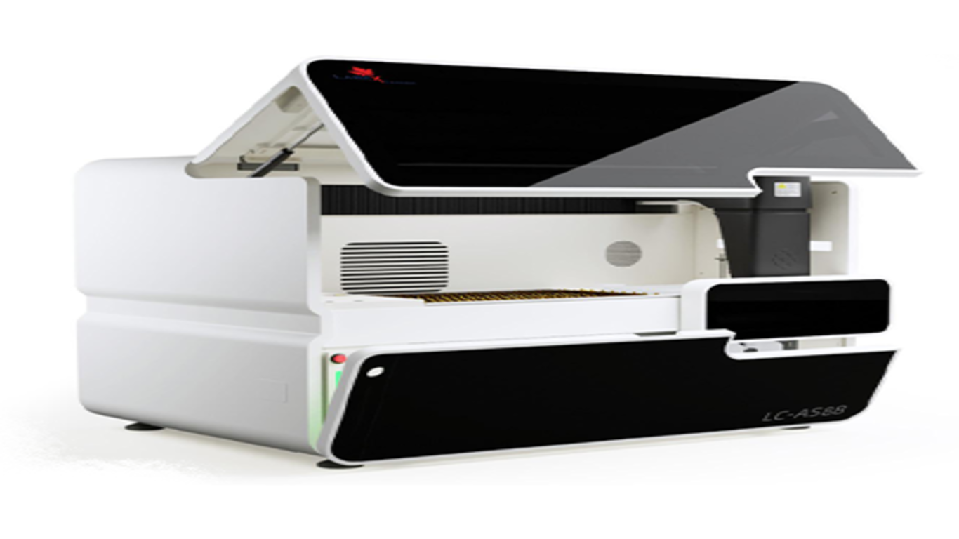
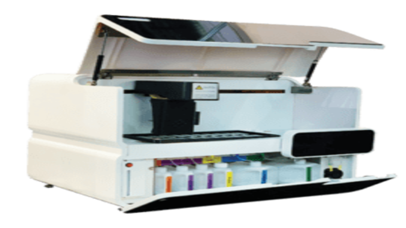
At Labex, we understand the importance of accurate and reliable cancer diagnosis. Our IHC staining services are designed to meet the highest standards of quality, with a focus on delivering precise results that guide clinical decision-making. Here’s why Labex is the preferred choice for IHC Stainer:
Experienced Pathologists and Technicians
Our team of experienced pathologists and technicians is dedicated to providing accurate IHC staining results. We use validated antibodies and standardized protocols to ensure consistency and reliability.
Comprehensive Marker Panel
We offer a comprehensive panel of IHC markers to address a wide range of diagnostic needs, from identifying tumor origin to assessing prognostic and predictive biomarkers. Our services are tailored to meet the specific requirements of each case.
State-of-the-Art Technology
Labex utilizes state-of-the-art staining equipment and image analysis software to deliver high-quality IHC results. Our technology ensures precise quantification of protein expression and supports the identification of subtle differences in staining patterns.
Commitment to Quality and Accuracy
Quality is at the core of everything we do. Our rigorous quality control procedures and attention to detail ensure that our IHC staining services meet the highest standards of accuracy.
Conclusion
IHC Stainer is crucial in cancer diagnosis, providing valuable information about tumor type, origin, and molecular characteristics. By enabling the detection of specific protein markers, IHC staining helps guide treatment decisions, assess prognosis, and support personalized medicine. Labex is committed to delivering high-quality IHC staining services that support accurate cancer diagnosis and improve patient outcomes.
If you need expert IHC Stainer services or would like to learn more about how we can help with your diagnostic needs, contact Labex today and let us be your trusted partner in cancer diagnostics.
